
Figure 6. Mtbp C-terminus functions as an inhibitor of Myc.
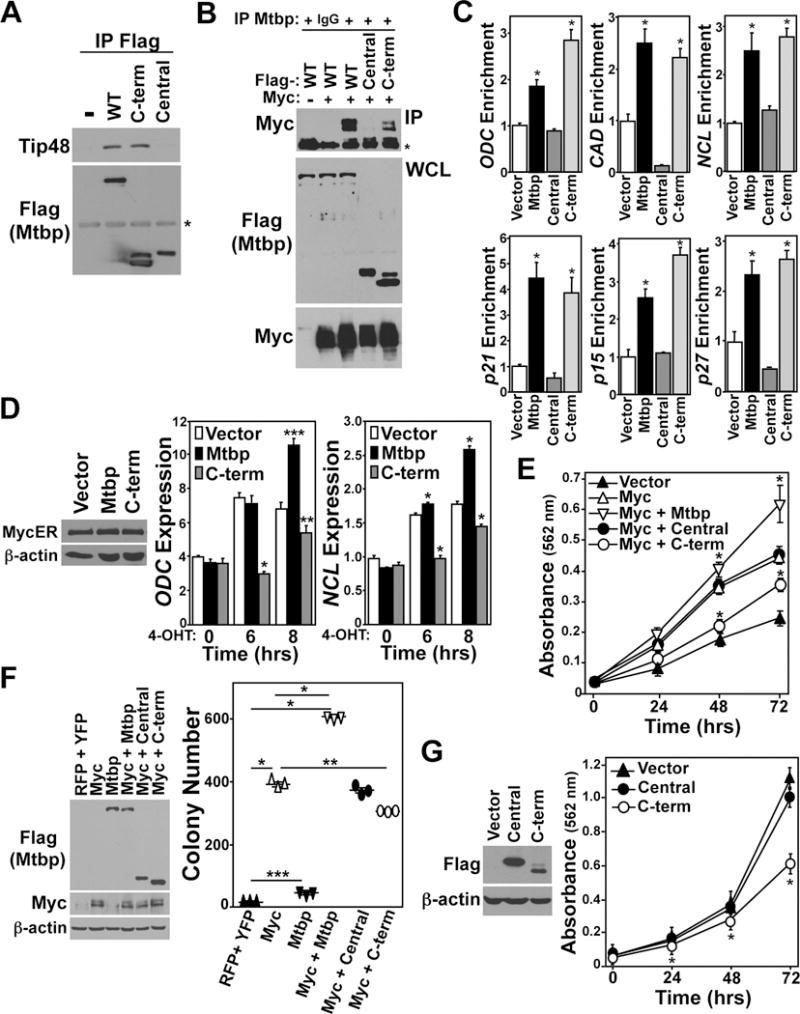
(A–C) 293T cells were transfected with vectors encoding Flag-tagged wild-type (WT) Mtbp, the C-terminal (597–894) or central domain (299–597) Mtbp mutants, or empty vector control (–) and for (B) a vector that did (+) or did not (−) encode Myc. (A) Anti-Flag immunoprecipitation of whole cell lysates were Western blotted for Flag and endogenous Tip48. Asterisk denotes location of immunoglobulin heavy chain. (B) Whole cell lysates (WCL) were Western blotted for Flag and Myc (below) and subjected to anti-Mtbp or IgG control IP and then Western blotted for Myc (top). (C) Following ChIP with anti-Flag, qRT-PCR was performed for the indicated promoter regions. Values are relative to their respective vector control and input DNA (*p≤0.01 compared to vector). (D) MycER expressing NIH3T3 cells were transfected with an empty vector or a vector encoding full-length Mtbp or the C-terminal Mtbp mutant. Western blots were performed. To activate MycER, 4-OHT was added to the cultures for the indicated time. qRT-PCR was performed in triplicate for the indicated Myc target genes and is expressed relative to β-actin levels (*p<0.001 vector vs. C-term or Mtbp, ***p=0.006 vector vs. Mtbp, **p=0.05 vector vs. C-term). (E) NIH3T3 cells transfected with empty vector or vectors encoding the indicated proteins were subjected to MTT assay at 24 hr intervals. (p<0.001 for Myc vs. Myc + Mtbp and Myc + C-term vs. Myc or Myc + Mtbp). (F) NIH3T3 cells infected with bicistronic retroviruses encoding the proteins indicated were subjected to soft agar colony assay. Colony number quantified after 10 days (*p<0.0001, **p=0.0004, ***p=0.0006). (G) HCC1806 cells expressing YFP alone (vector) or YFP with Flag-tagged central or C-terminal Mtbp mutants were Western blotted (left). Cells were also subjected to MTT assays at 24 hr intervals (*p<0.05 Vector vs. C-term). Error bars are standard deviation and p values determined by student’s t-tests.