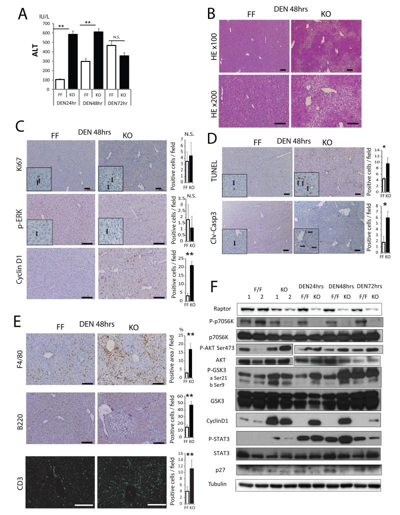
Figure 4. Loss of Raptor enhances hepatocyte death and inflammation after DEN challenge.
RaptorF/F (FF) and RaptorΔhep (KO) 8 weeks old mice were i.p. injected with 100 mg/Kg DEN and their sera and livers analyzed 24-72 hrs later. (A) Serum ALT after DEN injection (n= 3). (B,C) H&E, Ki67, p-ERK, and cyclin D1 staining of liver sections from FF and KO mice 48 hrs after DEN injection (n= 3). Bar graphs indicate frequencies of Ki67, p-ERK, and cyclin D1 positive cells. (D) Cell death in FF and KO livers was analyzed 48 hrs after DEN injection by TUNEL and immunostaining with the antibody against cleaved-caspase 3 (clv-casp 3). Quantitative analysis is shown in the bar graphs (n= 3). (E) Immune cell infiltration was analyzed 48 hrs after DEN injection by immunostaining with the indicated antibodies. F4/80 positive areas were quantified with Image J software. B220 and CD3 positive cells were counted (n= 3). (F) Immunoblot analysis of liver lysates collected after DEN injection. All the bar graphs in Figure 4 represent means +/− S.D. See also Figure S4.

