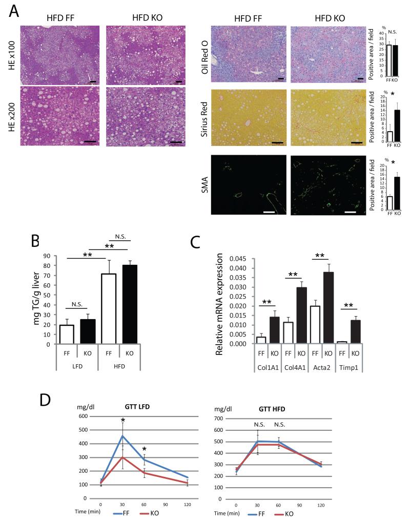
Figure 6. Hepatocyte Raptor ablation does not reduce hepatosteatosis and augments liver fibrosis in HFD fed mice.
RaptorF/F (FF) and RaptorΔhep (KO) mice were kept on HFD or LFD for 6 months. (A) Liver histology, lipid accumulation and fibrosis were analyzed by staining liver sections with H&E, oil red O, Sirius Red and α smooth muscle actin (SMA) antibody. The positive areas were quantified with Image J software and shown as bar graphs. (B) Liver triglyceride (TG) content in 7 months old mice kept on LFD or HFD. (C) The mRNA amounts of fibrogenic markers, collagen α1(I) (Col1A1), collagen α1(IV) (Col4A1), actin α (Acta), and TIMP1 (Timp1) in livers of 7 months old FF and KO mice were determined by real time qPCR. (D) Glucose tolerance tests were performed on above mice kept on LFD and HFD (n= 4). All the bar graphs in Figure 6 represent means +/− S.D. See also Figure S6.

