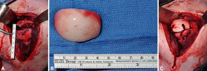
Fig. 6A–C.

(A) Intraoperative photograph of humeral head showing a large Hill–Sachs lesion indicated with a black arrowhead. (B) Fresh osteochondral humeral head allograft prepared for filling the Hill–Sachs defect. (C) The humeral head allograft is fit into final position affixed with headless compression titanium screws.
