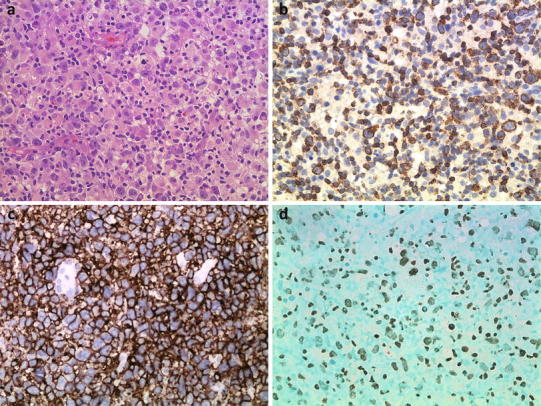
Fig. 1.

Pathological features of NK/TCL. Representative image of NK/TCL showing diffuse medium-sized to large and pleomorphic cell hyperplasia with a high mitotic rate (a H&E staining, ×400), positivity for CD3ε (b immunohistochemical staining, ×400), CD56 (c immunohistochemical staining, ×400), and EBER (d in situ hybridization, ×400)
