Fig. 4.
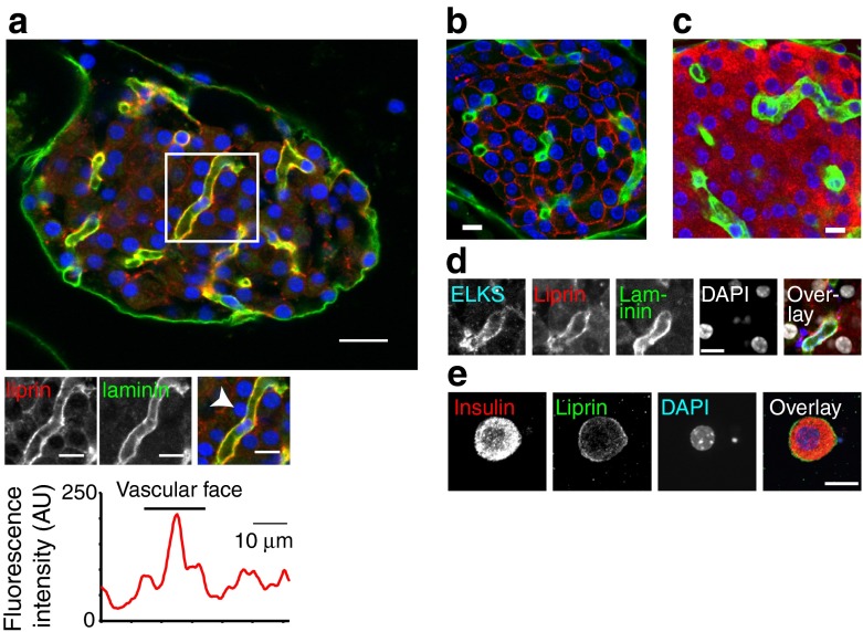
Presynaptic scaffold protein liprin is present in beta cells and is enriched at the vascular face. (a) Immunofluorescence image of islets shows laminin (green) as a marker of the basement membrane from the vascular endothelial cells and shown as low power, large images of a mouse whole islet (scale bar, 20 μm) and enlarged images of the regions bordered by the boxes (scale bar, 10 μm). Immunofluorescence of liprin (red) shows enrichment along the vasculature using a linescan around the cell (indicated by arrow) perimeter. (b, c) GLUT2 (red) and laminin (green) (b) and insulin (red) and laminin (green) (c) immunostaining show that GLUT2 and insulin are on the membrane of all cells in the islet core proving they are beta cells. (d) An isolated beta cell, with insulin immunostaining (red) and liprin (green) at the cell membrane. (e) Quadruple immunostaining shows that ELKS and liprin are enriched along the laminin-stained vasculature. Scale bar, 10 μm

