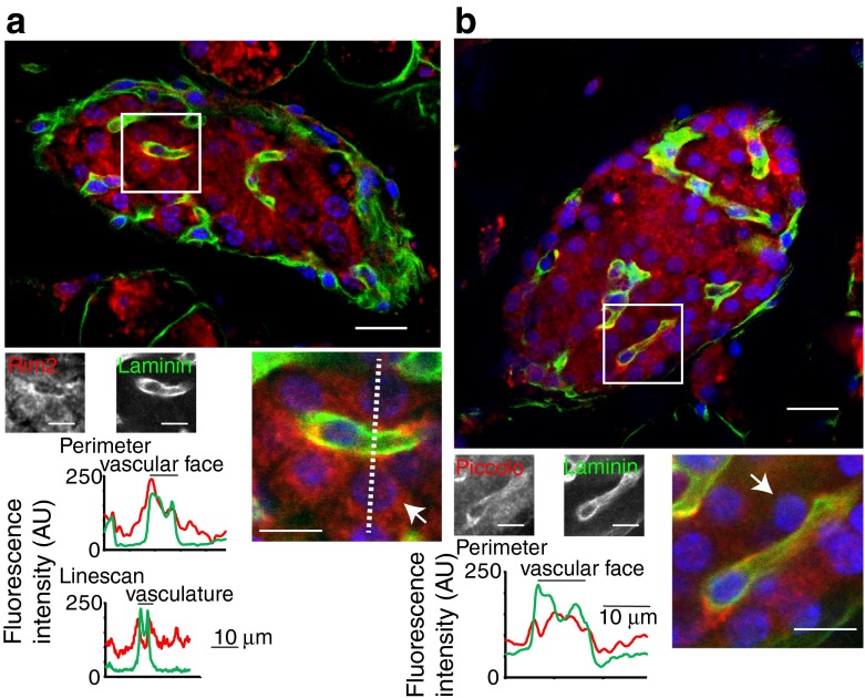
Fig. 5.
The presynaptic scaffold proteins Rim2 (a) and piccolo (b) are specifically enriched at the beta cell membrane that borders the vasculature. This enrichment is quantified in the histograms of the average fluorescence intensities either along a line drawn around the perimeter of the cells (indicated by arrows) or along a linescan (shown by the dotted line). Scale bars, 10 μm

