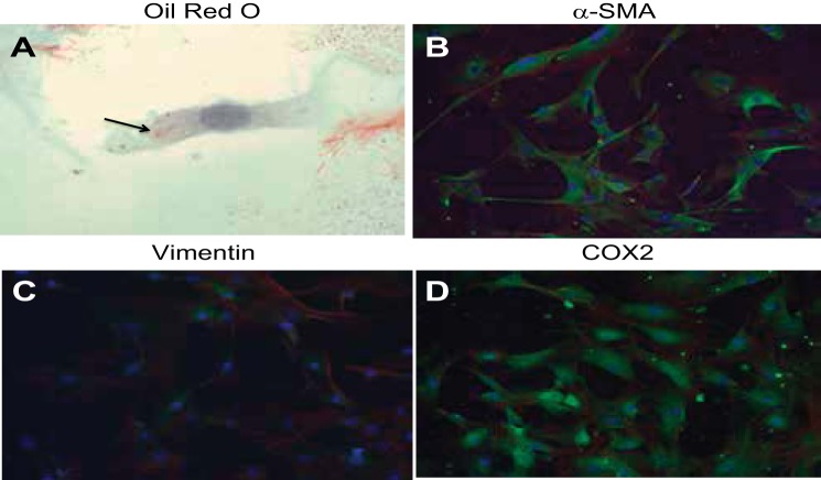
Fig. 1.
Characterization of mouse medullary interstitial cell (MMIC) primary cultures. Cells show high prevalence of oil red O-positive cytoplasmic inclusions (arrow) and elongated cytoplasmic extensions (A). MMICs show strong expression of α-smooth muscle actin (SMA; B, green) and cyclooxygenase (COX) 2 (D, green). MMICs also show trace vimentin expression (C, green). Green, green fluorescent protein (GFP)-label (antibody); blue, 4,6-diamidino-2-phenylindole (DAPI; nuclear); red, phalloidin (actin).

