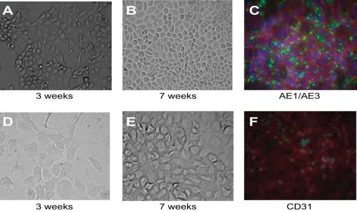
Fig. 11.
Cell phenotype differentiation by MPCs. A and B: progenitor cells were grown in epithelial growth medium (RenaLife Medium Complete Kit from LifeLine and α-MEM 1:1) for 7 wk. Progression of progenitor cell differentiated toward an epithelial phenotype, shown at 3 (A)- and 7-wk (B) growth periods. At 7 wk, progenitor cells grew in a cell monolayer with cell junctions (B) and positive expression of the pan-cytokeratin markers AE1/AE3 (C). Green, GFP label (AE1/AE3); red, phalloidin (actin); blue, DAPI (nuclear). Also shown is progression of progenitor cell differentiation toward an endothelial phenotype. Cells are shown at 3 (D)- and 7 (E and F)-wk growth periods. Progenitor cells show loose, rounded cell phenotype (D and E). Immunohistochemistry studies showed staining for the endothelial marker CD31 at 7 wk of growth (F). Green, GFP (CD31); red, phalloidin (actin); blue, DAPI (nuclear).

