Fig. 2.
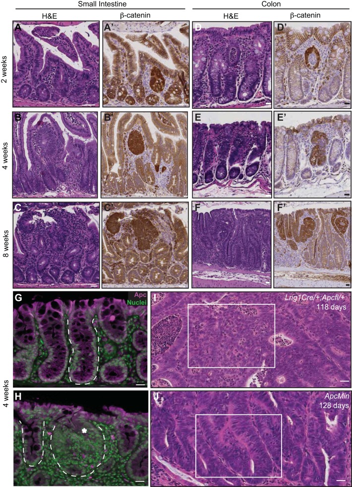
Histological progression of lesions in small intestine and colon of tamoxifen-treated Lrig1-Cre/+;Apcfl/+ mice. A–C: serial hematoxylin-eosin (H&E) staining and β-catenin immunoreactivity (brown) in small intestine 2, 4, and 8 wk postinduction. D–F: serial H&E staining and β-catenin immunoreactivity (brown) in colons 2, 4, and 8 wk postinduction. G: adenomatous polyposis coli (Apc, purple) immunoreactivity in a wild-type mouse colon. H: Apc immunoreactivity (purple) is not detected in a focal colonic microadenoma (asterisk) in a mouse 4 wk post-tamoxifen injection; however, Apc is detected in an adjacent normal-appearing crypt. In G and H, normal and transformed epithelial glands are outlined with a white dashed line. I and J: histological comparison of a typical tumor from an Lrig1-Cre/+;Apcfl/+ and an ApcMin mouse at 118 and 128 days of age, respectively. White box in I indicates high-grade dysplasia characterized by cribriform areas, with loss of nuclear polarity often observed in tumors from Lrig1-Cre/+;Apcfl/+ mice. White box in J indicates a low-grade dysplastic tubular adenoma with retained nuclear polarity characteristic of tumors from ApcMin mice. Scale bars, 25 μm.

