
Fig. 4.
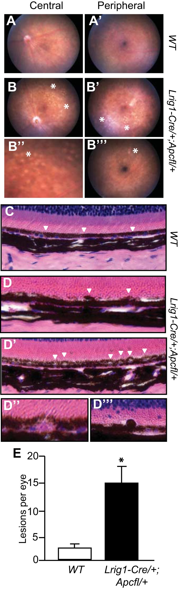
Lrig1-Cre/+;Apcfl/+ mice have defects in the retinal pigment epithelium (RPE). A and A′: fundus of a wild-type mouse with no obvious RPE defects in central or peripheral retina. B–B′′′: representative images of fundus abnormalities in an Lrig1-Cre/+;Apcfl/+ mouse. Areas of retinal depigmentation were found in the central retina (B and B′′; asterisks). Similar lesions were also present in the peripheral retina (B′ and B′′′). C: histological H&E analysis of a wild-type mouse showing normal outer retinal structure. RPE layer was evenly pigmented and had a smooth interface with the photoreceptor outer segment. D–D′′′: Lrig1-Cre/+;Apcfl/+ mice exhibited hyperpigmentation (arrowheads in D) and vacuolization of RPE cells (arrowheads in D′). Sub-RPE drusen-like deposits (D′′) and subretinal melanin-containing cell infiltration (D′′′) were also observed. E: Lrig1-Cre/+;Apcfl/+ mice had quantitatively more fundus lesions than their wild-type counterparts. Data are from Lrig1-Cre/+;Apcfl/+ mice >85 days postinduction and 5-mo-old wild-type mice. Values are means ± SE. *P < 0.01.