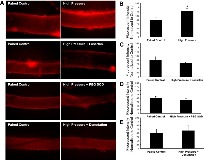
Fig. 8.
Superoxide levels in human adipose microvessels. A: representative fluorescent images of human adipose arterioles treated with 10 μmol/l dihydroethidium. Separate groups of vessels were exposed to high intraluminal pressure in the presence or absence of losartan (10−6 mol/l) or PEG-SOD (100 U/ml). In a fourth group of vessels, the endothelium was denuded by passing 20 ml of air through the lumen before exposure to high intraluminal pressure. To account for day-to-day and tissue-to-tissue variability, all experimental groups (right) were paired with a control vessel from the same tissue that only received dihydroethidium and was maintained at an intraluminal pressure of 60 mmHg (left). All images of vessel pairs from the same tissue have identical gain and threshold settings. B–E: vascular superoxide levels from each experimental group were normalized to the fluorescence intensity of the control vessel from the same surgical sample. Increased intraluminal pressure (n = 7 vessels) significantly increased dihydroethidium fluorescence in isolated human adipose arterioles compared with control vessels maintained at 60 mmHg from the same tissue (+53 ± 16% vs. control, P < 0.05; B). Treatment of vessels with losartan (10−6 mol/l, n = 6 vessels; C) or PEG-SOD (100 U/ml, n = 4 vessels; D) before exposure to an intraluminal pressure of 150 mmHg did not change hydroethidium fluorescence (−17 ± 3% and −13 ± 10% vs. control, respectively, P > 0.05). E: endothelial denudation (n = 4 vessels) prevented the pressure-induced increase in fluorescence (+17 ± 31% vs. control, P > 0.05) observed in untreated vessels. All values are plotted as means ± SE. Due to variations in the absolute fluorescent intensity of control vessels from tissue to tissue, results are plotted with the value of the control vessels normalized to 100. *Significant difference (P < 0.05) in high pressure vs. control.

