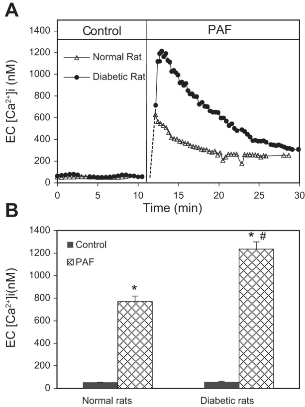
Fig. 2.

Diabetic vessels show higher Ca2+ responses to PAF than that in normal vessels. A: 2 representative experiments showing the differences in PAF-induced increases in endothelial intracellular Ca2+ concentration (EC [Ca2+]i) between normal and diabetic vessels. Endothelial [Ca2+]i in the diabetic vessel increased at a faster rate and reached a higher magnitude than that in a normal vessel. The time courses were derived from calcium imaging, and each point is the mean value of 12–18 regions of interest (endothelial cells) of the vessel wall. B: summary results of PAF-induced increases in endothelial [Ca2+]i in normal and diabetic vessels (n = 5/group). *Significant increases from control; #significant increases from normal vessel responses.
