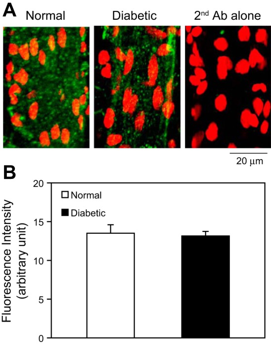
Fig. 3.

Immunostaining of PAF receptor in normal and diabetic mesenteric venules. A: representative confocal images of fluorescent immunostaining of the PAF receptor in normal and diabetic venules (green). The nuclei were labeled with DRAQ5 (red). The application of a secondary antibody (Ab) alone showed no significant green fluorescence. Each image is the maximum projection of ½ of the vessel. B: quantification of the mean fluorescence intensity (MFI) from normal and diabetic vessels (n = 3/group).
