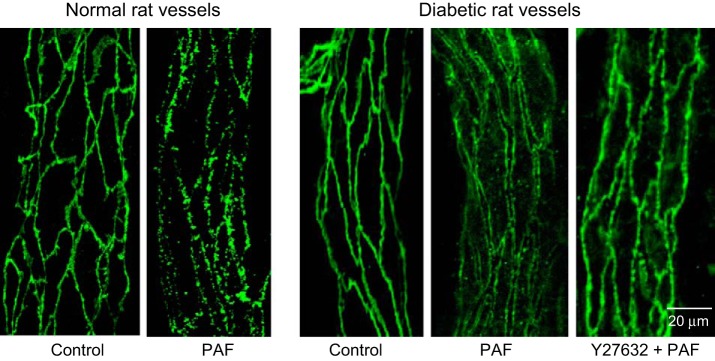
Fig. 5.

Vascular endothelial (VE)-cadherin staining under control and PAF-stimulated conditions in normal and diabetic rat vessels. VE-cadherin in normal rat vessels shows continuous distribution under control conditions and frequent breaks at the PAF-induced Lp peak. In diabetic vessels, VE-cadherin distribution under control conditions shows no difference from normal vessels but exhibits large gaps between endothelial cells with PAF stimulation. The application of Rho kinase inhibitor Y27632 replaces PAF-induced VE-cadherin separation with some breaks on a single profile distribution.
