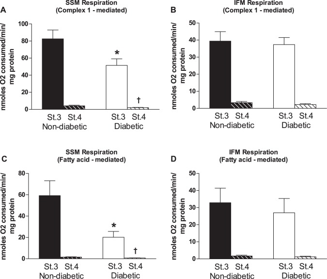
Fig. 2.
Mitochondrial subpopulation respiration rates. Cardiac mitochondrial subpopulations were isolated and respiration rates were measured using different substrates. Glutamate and malate were used as substrates to measure maximal complex I-mediated respiration [state (St) 3 and state 4] for nondiabetic and diabetic SSM (A; state 3, n = 28 patients without diabetes and n = 23 patients with diabetes; and state 4, n = 28 patients without diabetes and n = 22 patients with diabetes) and nondiabetic and diabetic IFM (B; state 3, n = 28 patients without diabetes and n = 23 patients with diabetes; and state 4, n = 28 patients without diabetes and n = 23 patients with diabetes). Palmitoylcarnitine and malate were used as substrates to measure maximal fatty acid-mediated respiration (state 3 and state 4) for nondiabetic and diabetic SSM (C; state 3, n = 16 patients without diabetes and n = 9 patients without diabetes; and state 4, n = 15 patients without diabetes and n = 9 patients with diabetes) and nondiabetic and diabetic IFM (D; state 3, n = 16 patients without diabetes and n = 13 patients with diabetes; and state 4, n = 16 patients without diabetes and n = 13 patients with diabetes). Values are means ± SE. Units are in nanomoles O2 consumed per minute per milligram of protein. Black bars represent patients without diabetes, and white bars represent patients with diabetes. *P ≤ 0.05, state 3 SSM nondiabetic vs. state 3 SSM diabetic; †P ≤ 0.05, state 4 SSM nondiabetic vs. state 4 SSM diabetic.

