
Figure 4. Transmission electron microscopy of purified virions binding to gB-GNP.
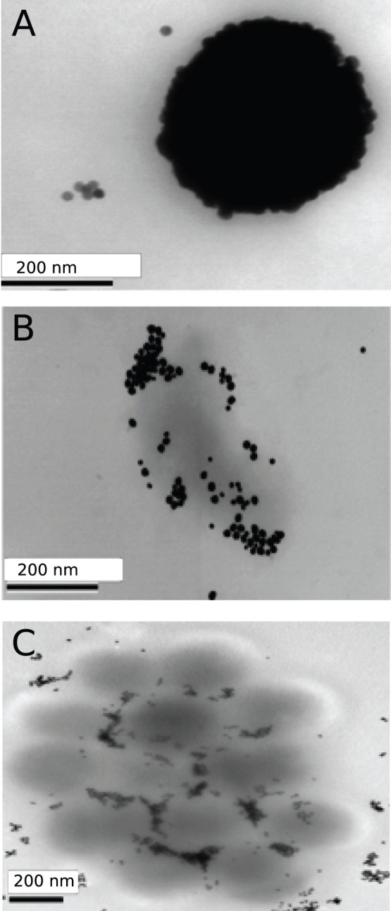
gB-GNPs conjugated to 100 ng antibody total (A) or 50 ng antibody total (B), (C) were added to purified virus particles and placed on a grid for electron microscopy. A single virion covered with gB-GNPs is shown (A), in comparison to a cluster of virions attaching to gB-GNPs (B), (C).