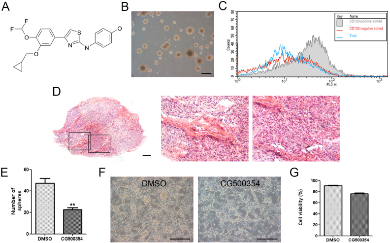
Figure 1. Characterization of human primary GBM-derived cells.
(A) Chemical structure of the small molecule CG500354. (B) Representative phase-contrast image of GBM-derived spheres in non-adherent culture conditions. Scale bar = 1 mm. (C) The GBM-derived cells were characterized by flow cytometry after sorting the CD133-positive cell population using magnetic-activated cell sorting. (D) A glioblastoma (left) from the GBM-derived cells in a xenograft mouse model has been characterized with pseudopalisading necrosis (middle) and endothelial proliferation (right) after hematoxylin and eosin staining. Scale bar = 400 μm. (E) CG500354 significantly reduced the number of spheres after 72 hours of exposure in a clonogenic assay. (F) Representative phase-contrast images of GBM-derived cells treated with DMSO or CG500354. Scale bar = 1 mm. (G) A trypan blue exclusion test was performed to calculate the number of viable cells compared to the total cell number in the GBM-derived cell population after 72 hours of treatment with CG500354 (3 μM) or DMSO. DMSO was used as a vehicle control.

