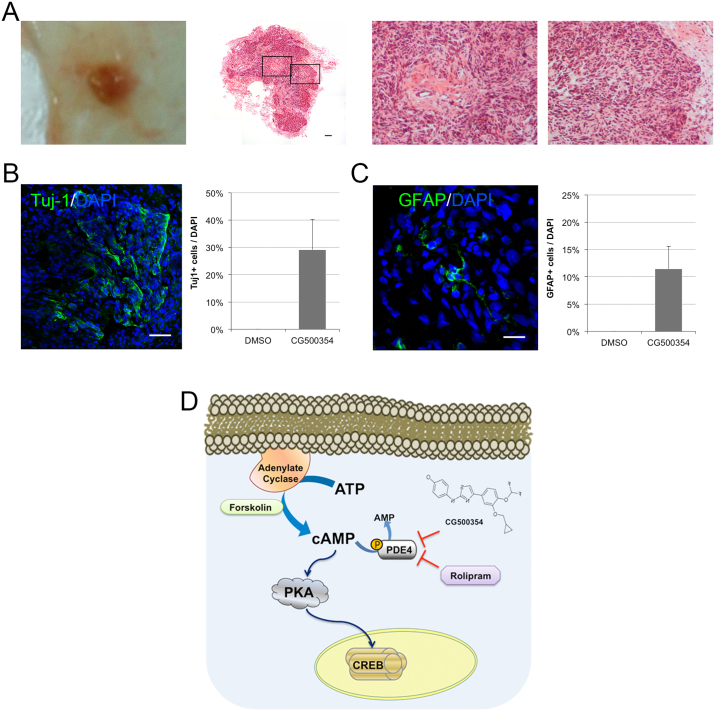
Figure 6. In vivo neural differentiation of human primary GBM-derived cells after xenotransplantation.
(A) After GBM tumor formation, tumors were isolated and stained with hematoxylin and eosin. A composited image (second from left) reveals an entire section of a tumor and two high-resolution images indicates pseudopalisading necrosis (second from right) and endothelial proliferation (right). (B–C) Representative immunochemical images of brain sections from GBM-derived tumors show that the cells inside of the tumors were forced to differentiate into Tuj1- and GFAP-expressing neural subtypes. (D) Schematic diagram of the mechanism of CG500354-triggered cAMP/CREB signaling pathway. Likewise, both of the mimetic substances Forskolin and Rolipram are involved in this signal transduction pathway.

