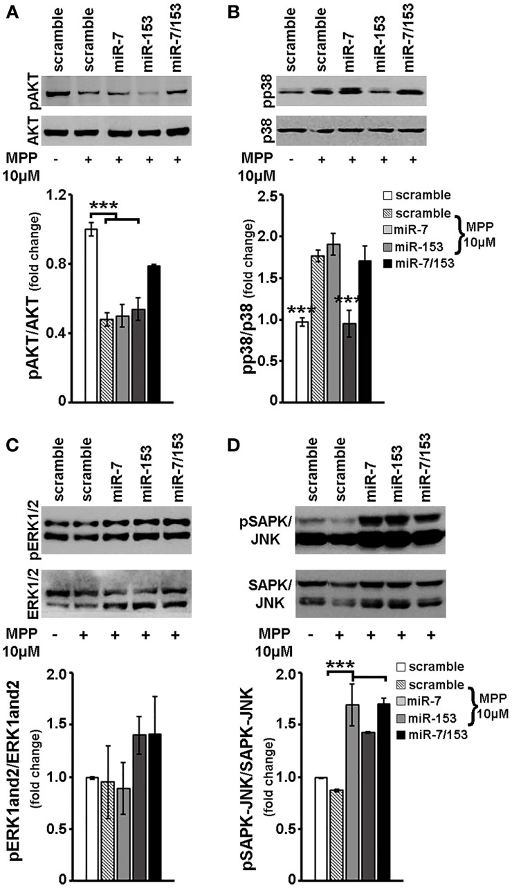
Figure 7.
Effect of miR-7 and/or mir-153 overexpression on AKT, p38, ERK-1/2, and SAPK/JNK signaling in MPP+-treated cortical neurons. Six to seven days old primary cortical neurons were transduced with adenoviral particles expressing scramble miR, miR-7, miR-153, or both miR-7/153. After 24 h, transduced neurons were exposed for additional 24 h to 10 μM of MPP+. Equal amounts of total protein from lysates of transduced cortical neurons cultured for 24 h in the presence of 10 μM MPP+ were analyzed on 10% SDS-PAGE and immunoblotted with antibodies specific for phosphorylated forms of AKT (A), p38 MAPK (B), ERK1/2 (C), and SAPK/JNK (D). To ensure equal loading membranes were re-probed against AKT, p38 MAPK, ERK1/2, and SAPK/JNK, respectively. Quantification of the results was performed by scanning densitometry. Bars in all the presented graphs depict mean ± s.e.m. Note that miR-7, miR-153, or miR-7/153 altered in an opposite way MPP+-induced changes in intracellular signaling cascades. ***P < 0.001.

