
Fig. 2.
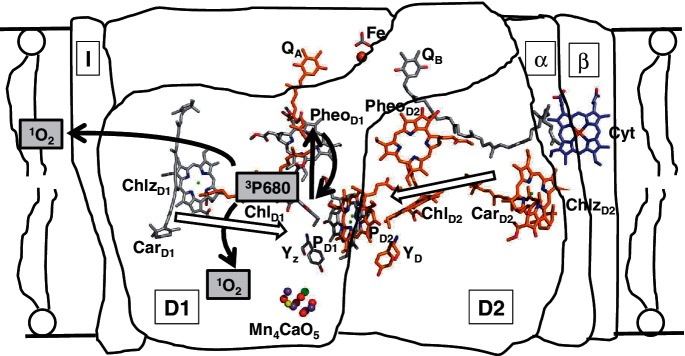
Schematic diagram of the electron transfer reactions occurring in the membrane-bound PSII RC and the formation of singlet oxygen at the site of 3P680. The purified complex (D1, D2, α and β subunits of Cyt b559 and the PsbI protein) has lost both of the secondary electron acceptors, QA and QB, the non-heme iron (Fe) and also the water-splitting Mn cluster, Mn4CaO5. The figure shows that if the triplet state of P680 is formed it will be quenched by ground state oxygen to form 1O2 which can damage either the pigment–protein complex or the lipid membrane. The cofactor arrangement in the RC complex shows the distance of the two β-carotenes from the four central Chl cofactors, based on Umena et al. (2011).