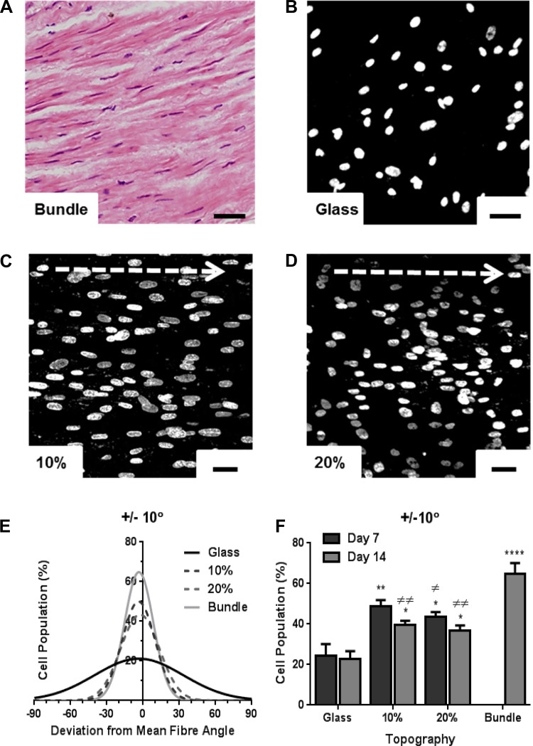
Fig. 2.
Human airway smooth muscle (HASM) cells orientate along scaffold fibers. Hematoxylin and eosin-stained immunohistological sections from airway smooth muscle (ASM) bundles were used to quantify cell directionality in situ (A). HASM cells (1.5 × 10⁵) were cultured on glass coverslips, the 10%, or 20% aligned scaffolds for 7 or 14 days prior to fixation. Nuclei were visualized by Hoechst staining (blue) and nuclei angles were used as reference to cell directionality. Deviation of individual cell nuclei from average fiber orientation was determined, and % cell population range was plotted. Representative images of HASM nuclei cultured on glass, 10%, or 20% aligned scaffolds at day 7 are shown in B, C, and D, respectively. E shows distribution of cell alignment on all 3 in vitro topographies and in ASM bundles. F shows % cell population within ±10° fiber orientation at day 7 and day 14 (means ± SE, HASM cells cultured on 3 independently electrospun scaffolds). Statistical significance is indicated as *P < 0.05, **P < 0.01, and ****P < 0.0001 (vs. glass), or ≠P < 0.05, and ≠≠P < 0.01 (vs. bundle), one-way ANOVA, Tukey's posttest. Scale bar indicates 40 μm. Arrow indicates orientation of scaffold fibers.

