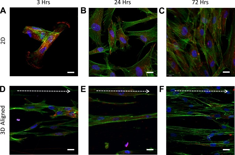
Fig. 4.
HASM cells show a reduction in intracellular-stress fibers when cultured on aligned fiber topographies. HASM cells (1.5 × 10⁵) were cultured on glass coverslips or 10% aligned scaffolds for 3, 24, or 72 h prior to fixation. Cells were immunostained for the focal adhesion protein vinculin (red), with F-actin stained with phalloidin (green) and nuclei with Hoechst (blue). Representative images of HASM cultured on glass coverslips for 3, 24, and 72 h, and 10% aligned scaffolds for 3, 24, or 72 h are shown in A, B, C, and D, E, F, respectively. Scale bar indicates 20 μm. Arrow indicates orientation of scaffold fibers.

