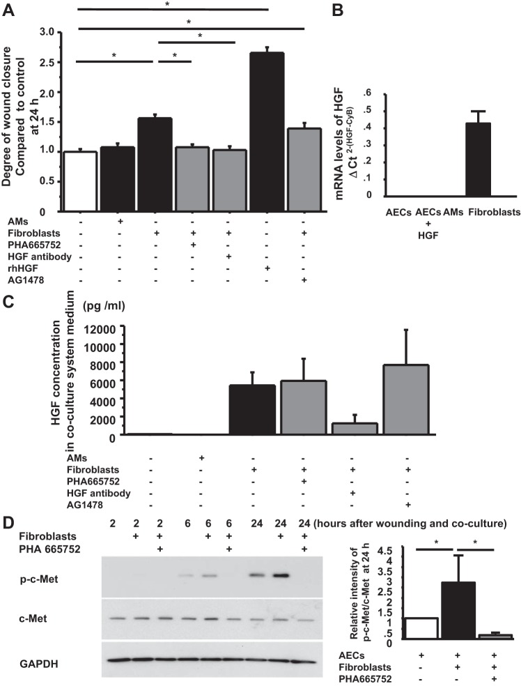
Fig. 3.
FBs promote wound closure though HGF/c-Met signaling. A: phase-contrast pictures were taken from marked wound areas at 0 and 24 h after wounding to compare the degree of wound closure among treatment groups: wounded AEC monolayer without coculture, wounded AEC monolayer cocultured with AMs, with FBs, with FBs + PHA 665752, with FBs + anti-HGF antibody, with HGF (as a positive control) and with FBs + AG 1478. The degree of wound closure was analyzed at 24 h after wounding. Values were means ± SE for 6 independent experiments. *P < 0.05. B: mRNA levels of HGF were measured by real-time PCR. These levels were normalized to the constitutive probe cyclophilin B (CyB). Values are means ± SE. C: HGF protein concentration in coculture media at 48 h after wounding was measured by ELISA. Values were means ± SE for 6 independent experiments. D, left: human AEC monolayers with wounds were cultured without FBs, with FBs, and with FBs + PHA 665752. The AECs were harvested at 2, 6, and 24 h after wounding, and protein levels of phospho-c-Met and c-Met normalized by GAPDH were measured by immunoblotting. This is a representative blot of 3 reproducible experiments. Right: relative intensity of p-c-Met/c-Met at 24 h by Western blotting analyzed by Image J from 3 independent experiments. Values are means ± SE for 3 independent experiments. *P < 0.05.

