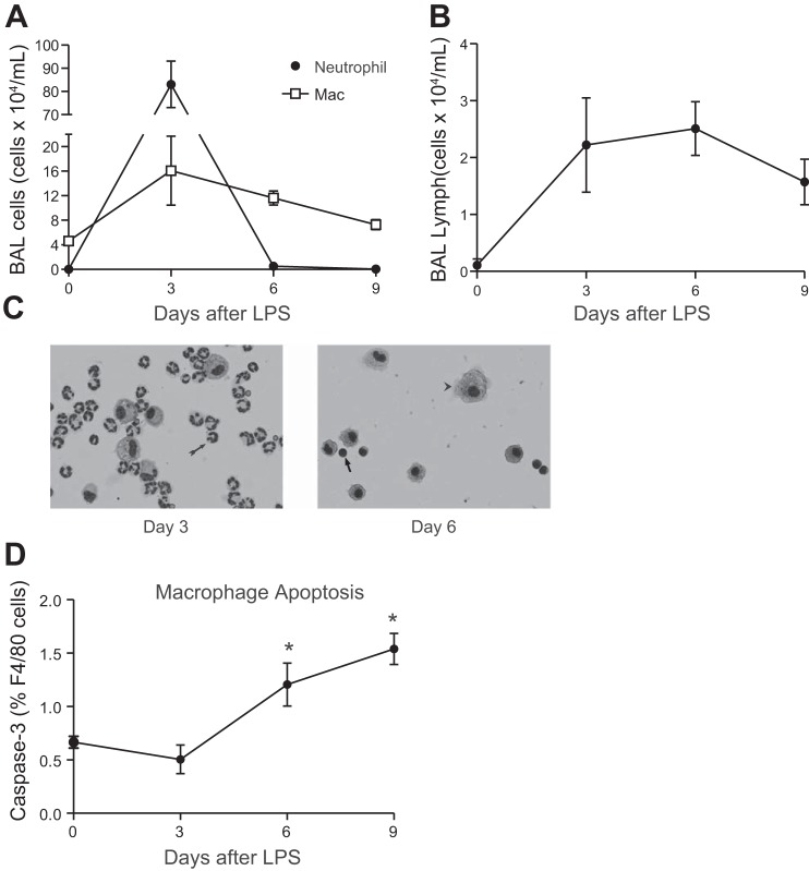
Fig. 1.
Cellular populations in bronchoalveolar lavage (BAL) fluid following intratracheal LPS. C57BL/6 mice were instilled with 20 μg of intratracheal LPS. Leukocytes were quantified using a hemacytometer. Cell differentials were determined by light microscopy of Wright-Giemsa-stained cytospin specimens or flow cytometry. A: total neutrophil and macrophage (Mac) counts; n = 4–8 mice per time point; 2 replicates. B: T-lymphocyte counts. T lymphocytes were identified by flow cytometry [side scatter angle (SSC) low, CD3+]; n ≥ 6 mice per time point; 2 replicates. C: representative Wright-Giemsa-stained cytospin specimens of BAL fluid (×20). Neutrophils (arrow, day 3), macrophages (arrowhead, day 6), and lymphocytes (arrow, day 6) are demonstrated. D: percentage of alveolar macrophages (AMΦs) expressing activated caspase-3 as quantified by flow cytometry; n = 4 mice per time point; 1 replicate. *P < 0.05.

