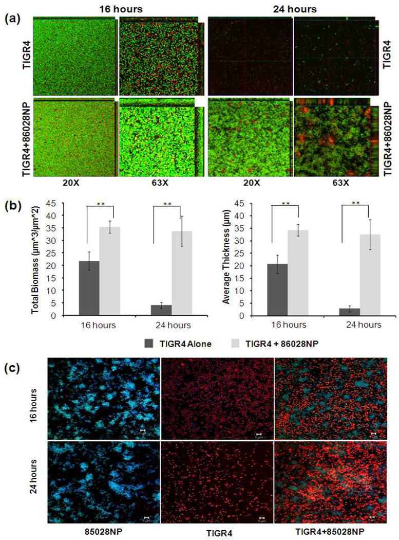
Fig. 2.
NTHi promoted pneumococcal biofilm formation and survival. (a). Biofilms grown in chambered cover glasses were visualized by CLSM following the bacterial live/dead staining process. The images are representatives of a single slice of each biofilm. (b). Total biomasses and average thicknesses of biofilm were analysed using COMSTAT. Mixed biofilms had more biomasses and were thicker than biofilms of TIGR4 alone at both time points. The values of biomass and thickness represented the averages of 5 Z-series images taken from different field of views, respectively. **: P<0.01. (c). TIGR4 and 86028NP cells in the biofilms were identified by fluorescent Gram staining followed by confocal laser scanning microscopy. 86028NP cells (Gram negative) were blue (live) or green (dead). TIGR4 cells (gram positive) were orange. The bar of microscopic image was 10µm.

