Fig. 5.
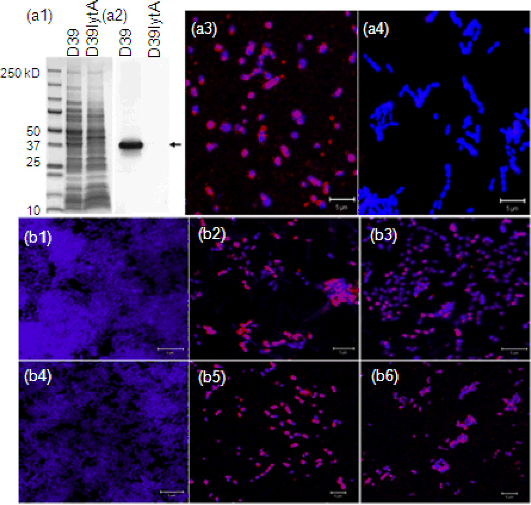
Expression and binding of pneumococcal autolysin in biofilm were decreased in the presence of NTHi. Pneumococcal LytA proteins produced (red) in the biofilm were detected by immunostaining with anti-LytA serum followed by confocal laser scanning microscopy. Biofilms were counterstained with DAPI (purple). a1–4: binding–specificities of anti-LytA serum were confirmed by western blot and immunostaining with D39 and its isogenic mutant D39lytA-. a1, results of SDS-PAGE; a2, results of western blot; a3, immunostaining of D39 biofilm; a4, immunostaining of D39lytA- biofilm. b1–6: results of immunostaining. b1, 16 hour biofilm of 86028NP; b2, 16 hour biofilm of TIGR4; b3, 16 hour biofilm of TIGR4 mixed with 86028NP; b4, 24 hour biofilm of 86028NP; b5, 24 hour biofilm of TIGR4; b6, 24 hour biofilm of TIGR4 mixed with 86028NP. At both time points, there were more LytA proteins in the biofilm of TIGR4 alone than the mixed biofilm cultures. The bar of microscopic image was 10 µm.

