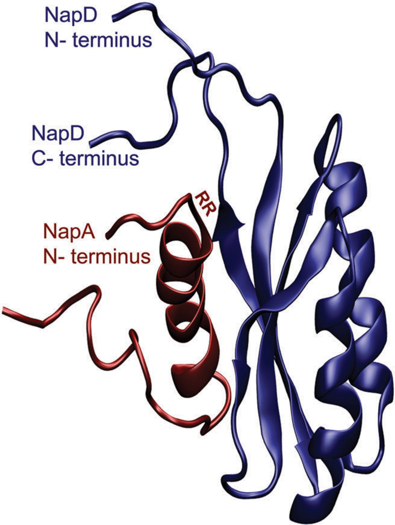
Fig. 15.
The protein structure of E. coli NapD in complex with the N-terminus of NapA. The protein backbone, of NapD (blue) and NapA (red), is displayed as a ribbon. The location of the two arginine residues (RR), for which the TAT leader sequence is named, is on the N-terminus of NapA. The figure the NMR solution structure (PDB ID: 2PQ4 was created using VMD (version 1.8.7) software.

