
Figure 11.
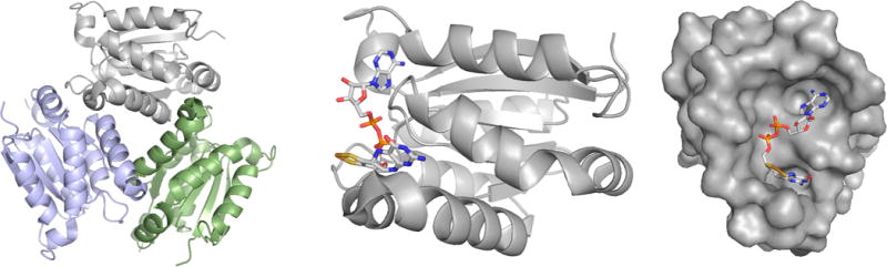
The structure of MogA homologues. Left, the common trimeric structure of the proteins, as illustrated by the G domain of human gephyrin (PDB 1JLJ). Center, a single subunit of the A. thaliana Cnx1-G domain (shown in approximately the same orientation as the gray subunit of the trimer at left), in complex with its product, the adenylylated pyranopterin cofactor (MPT·AMP). The structure is of a fully functional S583A mutant (PDB 1UUY). Right, the surface of the A. thaliana G domain showing the deep crevice in which the product binds (the orientation is rotated 90° about the vertical as compared to that seen at center).