
Figure 19.
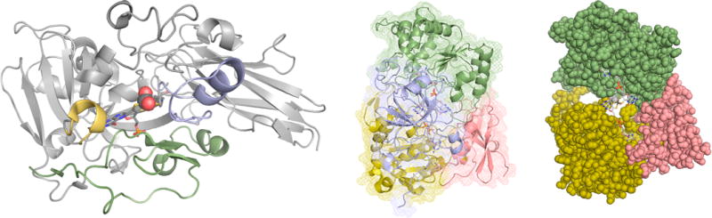
Protein structure and cofactor insertion in sulfite oxidase and TMAO reductase. Left, the backbone trace of the A. thaliana sulfite oxidase (PDB 1OGP). The structural elements shown in yellow, blue, and green are in positions that could allow them to transiently swing away in a largely folded apoprotein to accommodate the incoming cofactor. Center, the structure of S. massilia TMAO reductase (PDB 1TMO), as seen from the back of the protein opposite the substrate access funnel, with Domains I-IV in red, yellow, green, and blue, respectively. Right, a space-filling representation with Domain IV removed, exposing the enzyme’s molybdenum center from the back.