
Figure 2.
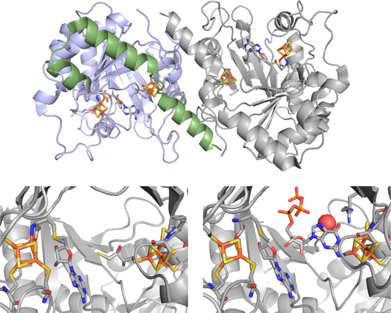
The structure of MoaA from S. aureus. Top, the structure of the dimeric enzyme, with the N- and C-terminal domains of the subunit on the left shaded in blue and green, respectively. Bottom left, a close-up of the active site with S-adenosylmethionine bound to the N-terminal [4Fe-4S] cluster and dithiothreitol bound to the C-terminal [4Fe-4S] (PDB 1TV8). Bottom right, a close-up of the active site of GTP bound to the C-terminal cluster, and methionine bound at the N-terminal cluster; 5′deoxyadenosine is also present in the active site (PDB 2FB3).