
Figure 20.
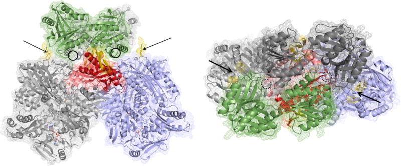
A model for the interaction between R. capsulatus xanthine dehydrogenase (PDB 2W3R) and the B. halodurans XdhC homologue (PDB 3ON5). The xanthine dehydrogenase has its two αβ protomers colored light gray and blue, with the previously identified interaction motif of each protomer in red. The redox-active centers of the dehydrogenase are rendered as CPK-colored spheres. The XdhC homologue is rendered with its subunits in dark gray and green. The insets present in the bacterial members of the xanthine oxidase family (but absent in eukaryotic members) are indicated in yellow. The general locations of the cofactor binding sites in the latter are indicated by the circles. The orientation at right is rotated 90° about the horizontal relative to that at left.