
Figure 38.
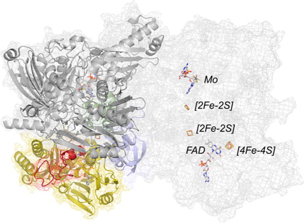
The structure of 4-hydroxybenzoyl-CoA reductase (PDB 1RM6). The protomer at left is color coded with the two domains of the iron–sulfur-containing subunit in blue and green, the FAD-containing subunit in yellow (with the [4Fe-4S]-containing inset in red), and the molybdenum-containing subunit in gray. The protomer at right is shown in mesh so as to more clearly illustrate the disposition of the several redox-active centers with respect to one another (rendered in CPK coloring).