
Figure 45.
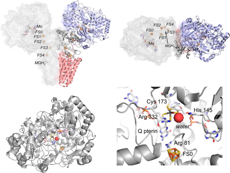
The structure of the PsrABC polysulfide reductase from T. thermophilus (PDB 2VPW). Top, the overall organization of the subunits in the (αβγ)2 oligomer. One protomer at left is in gray and shown in mesh to illustrate the disposition of the redox-active centers within the protomer, the other has PsrA, PsrB, and PsrC in blue, dark gray, and red, respectively. The orientation at right is rotated 90° about the horizontal compared to that at left. Bottom left, the PsrA subunit, looking down the solvent access channel to the active site. Bottom right, the active site molybdenum center, with Cys 173 coordinating the molybdenum and Arg 81 intervening between the Q pterin and FS0. Arg 332 and His 145 H-bond to a bound water.