
Figure 57.
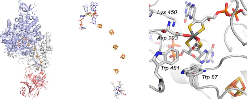
The structure of ethylbenzene dehydrogenase from Aromatoleum aromaticum (PDB 2IVF). Left, the overall structure of the trimeric enzyme, with the α, β, and γ subunits in blue, gray, and red, respectively. Center, an enlargement of the enzyme’s electron transfer chain, with the molybdenum center at top and the heme at bottom. Right, the active site of the enzyme, with residues referred to in the text indicated.