
Figure 9.
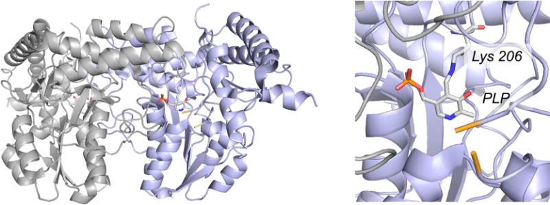
The structure of the E. coli cysteine desulfurase IscS (PDB 1P3W). Right, the overall fold of the dimer, with the two subunits shown in gray and blue and the pyridoxal phosphate rendered in CPK colors. Left, a close-up of the active site, showing the pyridoxal phosphate present as a Schiff base with Lys 206. Residues 327 and 334, which delimit the unresolved residues 328–333 (including Cys 328 that is made of a persulfide in the course of the desulfurase reaction), are shown in yellow.