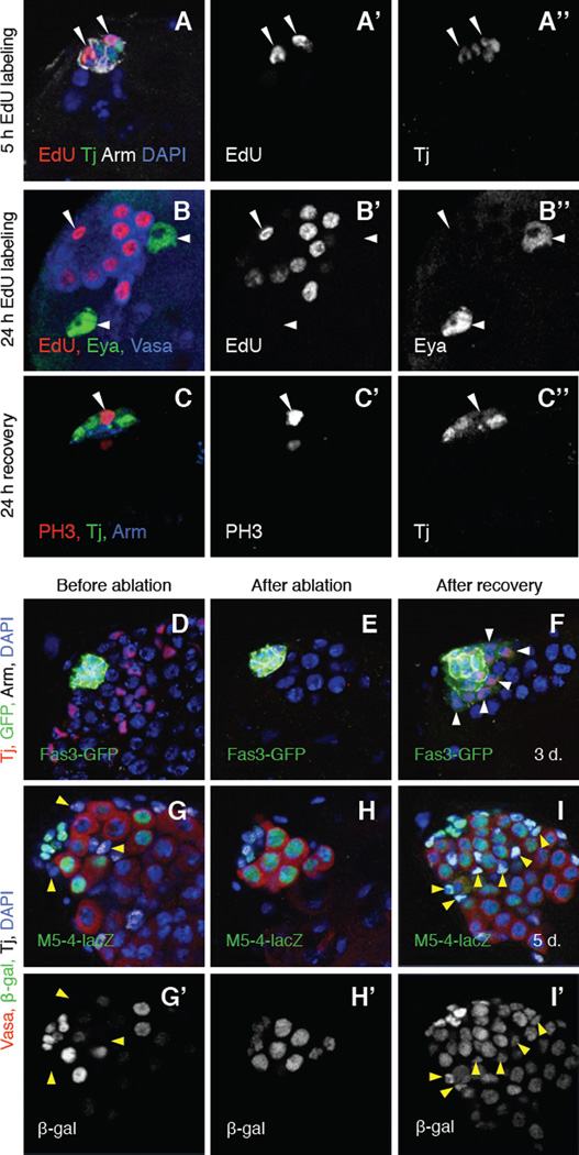
Figure 2. After complete genetic ablation of CySCs, hub cells exit quiescence and convert into CySC-like cells.
(A–I) Single confocal sections through the apex of c587-Gal4>grim testes. Nuclei are counterstained with DAPI (A, D–I, blue). (A–A”) Testis labeled with S-phase marker EdU (red and A’), anti-Tj (green and A”; hub cells) and anti-Arm (white; hub cells). After CySC ablation and 5 hours of recovery with EdU labeling, some hub cells incorporate EdU, indicating that they are no longer quiescent (arrowheads). (B-B”) Testis labeled with EdU (red and B’), anti-Eya (green and B”; late cyst cells), and Vasa (blue; germ cells). After CySC ablation and 24 hours of recovery with EdU labeling, EdU is incorporated into some somatic cells in or adjacent to the hub (one hub cell shown, arrowhead) and into some germ cells (Vasa-positive) but not into any Eya-positive cyst cells (two shown, arrows). (C–C”) Testis labeled with the mitotic marker anti-PH3 (red and C), anti-Tj (green and C”), and anti-Arm (blue). After CySC ablation and 24 hours of recovery, some hub cells express PH3 (arrowhead), indicating that they have entered mitosis. Tj disperses throughout the cytoplasm of mitotic cells. (D–F) Testes immunostained with anti-Tj (red), anti-GFP (green, alone in insets), and anti-Arm (white). GFP expression is driven by the Fas3 promoter. (D) Before and (E) after ablation, GFP is expressed throughout the hub but not in any cells outside the hub. (F) After 3 days of recovery, GFP is found in the hub and in regenerating CySCs (Tj -positive/Arm-negative cells outside the hub; arrows) in 81% of testes (n = 22/27). (G–I) Testes immunostained with anti-Vasa (red), anti-β-Gal (green and G’-I’), and anti-Tj (white). β-Gal expression is driven by M5-4-lacZ. (G–G’) Before and (H–H’) after ablation, M5-4-lacZ marks hub cells and early germ cells but not CySCs or cyst cells (arrows). (I–I’) After 5 days of recovery, β-Gal is found in hub cells, germ cells, and in regenerating CySCs and early cyst cells (Tj-positive/Arm-negative cells outside the hub; arrows). This is likely to reflect continued transcription, rather than perdurance, of the marker. Scale bars, 20µm. See also Figure S2 and Tables S2 and S3.

