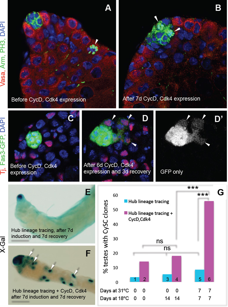
Figure 3. Ectopic CycD-Cdk4 expression in the hub promotes hub cell proliferation and conversion to CySCs.
(A–D) Single confocal sections through the apex of testes expressing CycD and Cdk4 in hub cells, immunostained as indicated. Nuclei are counterstained with DAPI (blue). (A –B) E132-Gal4>CycD, Cdk4 testes immunostained with anti-Vasa (red; germ cells), anti-Arm (green at cell cortex; hub cells), and anti-PH3 (green; mitotic nuclei). (A) Before CycD-Cdk4 induction (18°C), PH3-positive cells (arrow) are found outside but never inside the hub (n = 127 testes). (B) After CycD-Cdk4 expression (7 d, 31°C), 42% of testes (n = 35/83) contain PH3-positive hub cells (two shown, arrowheads). (C–D) E132-Gal4>CycD, Cdk4 testes co-expressing Fas3-GFP (marking hub cells), immunostained with anti-Tj (red; hub cells, CySCs and early cyst cells) and anti-GFP (green). (C) Before CycD-Cdk4 expression (18°C), GFP expression is restricted to the hub. (D-D’) After CycD-Cdk4 expression (6 d, 31°C) and recovery (3 d, 18°C), GFP is observed in Tj-positive cells outside the hub (arrows) in 23% of testes (n = 7/30). (E, F) X-gal staining revealing lineage traced hub cells (outlined) in the absence or presence of ectopic CycD-Cdk4. Testes contain either (E) marked hubs (example is from a control male, genotype: E132-gal4;;UAS-Flp/Act5c>Stop>lacZ), or (F) marked hubs and marked CySCs and their descendants (F, arrows), referred to herein as “CySC clones” (example is from an experimental male, genotype: E132-gal4; UAS-CycD, UAS-Cdk4; UAS-Flp/Act5c>Stop>lacZ). (G) Bar graph showing that flies expressing CycD-Cdk4 in hub cells have significantly more CySC clones than age-matched uninduced flies of the same genotype (bars 4 vs. 6), and than flies lacking ectopic CycD-Cdk4 that are processed in parallel (bars 5 vs. 6). The low percentage of CySC clones in testes from newly eclosed males represents background levels of labeling; this does not change significantly over time (bars 1,3,5). ns, not significant. *** P value < 0.0001 (two-tailed Fisher’s exact test). Scale bars, 20 µm (A–D) or 100 µm (E–F). See also Table S4.

