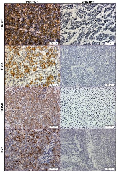
Figure 1. Activation of mTOR pathway in MCC tissues.
Representative positive immunohistochemical staining of P-4E-BP-1, P-S6K, P-mTOR, and MCV in sections from MCC TMA (left column). Representative negative immunohistochemical staining of P-4E-BP-1, P-S6K, P-mTOR, and MCV in sections from MCC TMA (right column)

