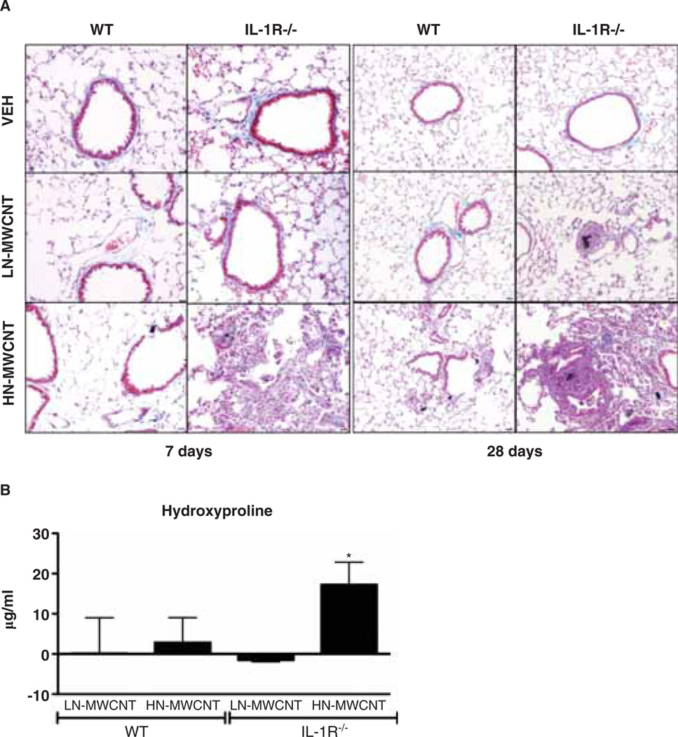
Figure 4.
Collagen deposition is augmented in the lungs of MWCNT-treated mice. Histopathology of lungs of IL-1R−/− mice exposed to MWCNT displays granuloma-like lesions and fibrotic tissue at 7 and 28 days post exposure. The WT or IL-1R−/− mice were euthanised at days 7 and 28 post exposure. After perfusion, the lung tissue from mice instilled with DM only or 50 mg LN- or HN-MWCNT was fixed in paraformaldehyde and imbedded in paraffin. Tissue sections that were stained with Gomori’s Trichrome demonstrate collagen-rich granulomas and surrounding fibrotic tissue in lungs of both WT and IL-1R−/− mice exposed with HN-MWCNT, but not DM (vehicle) control or LN-MWCNT-exposed mice (A). Collagen deposition in the lungs of WT or IL- 1R−/− mice following instillation with LN-, HN-MWCNT was assessed by hydroxyproline assay (n = 3–5 mice/group). The data were normalised by subtracting the mean background from the vehicle-treated mice, from the hydroxyproline levels of the particle-treated animals. Values are means ± SEM; * p < 0.05 compared to vehicle controls (B)

