Figure 2.

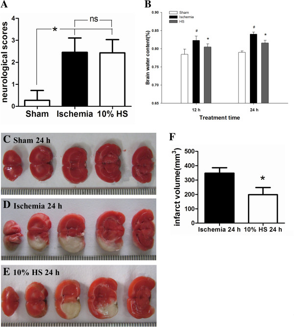
The effect of 10% HS on BWC and infarct size. Bar graph A shows that the neurological score shows no difference between the ischemia group and the 10% HS group (ns: P >0.05, n = 7 per group). Bar graph B shows that the percentage of BWC is significantly increased in the ipsilateral ischemic hemisphere at 12 h and 24 h following MCAO when compared with corresponding controls (sham-operated) (# P <0.05). However, it is significantly decreased in the ipsilateral ischemic hemisphere in 10% HS groups as compared with corresponding ischemic group (* P <0.05, n = 7 per group). TTC staining images (C-E) shown are from three representative experiments. The white area represents the infarct brain, and the red area represents the normal brain. Bar graph F shows the infarct volume is significantly decreased in 10% HS group when compared with ischemia group (* P <0.05, n = 6 per group). Note 10% HS could reduce infarct size effectively. Scale bars: (C-E), 1 mm per compartment. BWC, brain water content; HS, hypertonic saline; MCAO, middle cerebral artery occlusion; TTC, 2, 3, 5-triphenyltetrazolium chloride.
