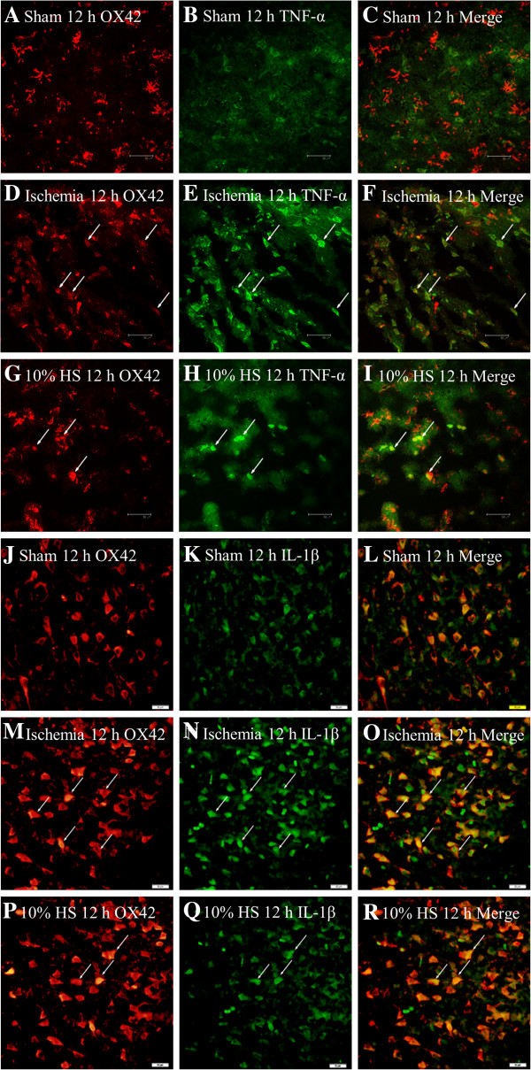
Figure 4.

Ten percent HS reduced the TNF-α and IL-1β expression in microglia-located peri-ischemic brain tissue. Confocal images showing the distribution of OX42 labeled microglia (A, D, G, J, M, P, red), TNF-α (B, E, H, green) and IL-1β (K, N, Q, green) in peri-ischemic brain tissue at 12 h after MCAO and the corresponding control rats. OX42 labeling overlapping TNF-α immunofluorescence can be seen in C, F and I, OX42 labeling overlaps IL-1β immunofluorescence in L, O and R. Note that TNF-α and IL-1β expression in microglia cells (arrows) is markedly enhanced following MCAO. However, after treatment with 10% HS, they are noticeably reduced. Scale bars: (A–R), 50 μm. HS, hypertonic saline; IL-1β, interleukin- 1beta; MCAO, middle cerebral artery occlusion; TNF-α, tumor necrosis factor-alpha.
