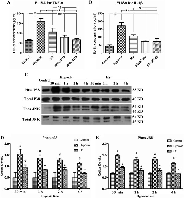
Figure 6.

HS attenuated hypoxia-induced TNF-α and IL-1β expression by inhibiting the activation of p38 and JNK signaling pathways. Panels A and B show levels of TNF-α (A) and IL-1β (B) protein in cultured medium from microglia decreased significantly after treatment with 100 mM HS, SB203580 and SP600125, as compared with the hypoxia group (*P < 0.05, **P < 0.01 ). Panel C shows p38, JNK phosphorylation and total p38, JNK immunoreactive bands. Bar graphs D and E show the optical density of phosphorylated p38 (38 kDa) and JNK (46 kDa, 54 kDa) immunoreactive bands are significantly increased at 30 minutes, 1 h, 2 h and 4 h following hypoxia, respectively (#P < 0.05). However, after treatment with 100 mM HS, the optical density of phosphorylated p38 and JNK is significantly decreased as compared with the corresponding hypoxia groups (*P < 0.05). ns: non-significant, P > 0.05. HS, hypertonic saline; IL-1β, interleukin- 1beta; TNF-α, tumor necrosis factor-alpha.
