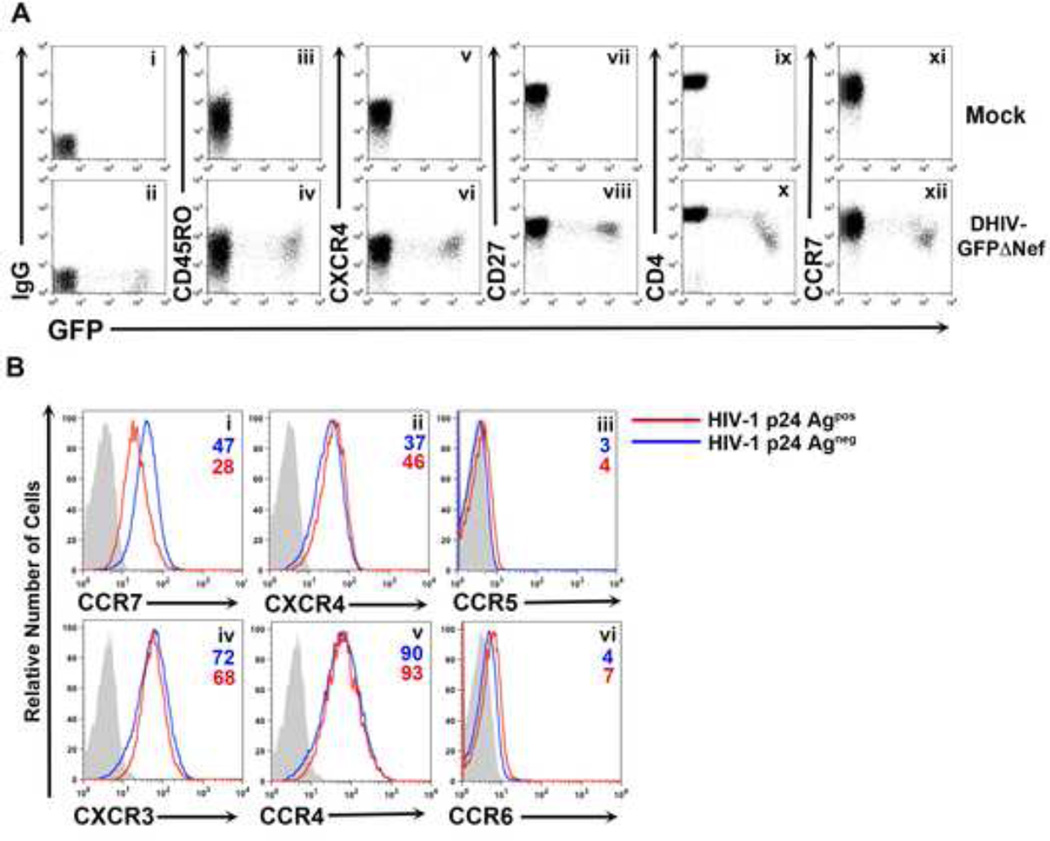
Figure 1. HIV-1 downregulates the chemokine receptor CCR7 from the surface of infected primary CD4+ T cells.
A) Surface levels of CD45RO (iii, iv), CXCR4 (v, vi), CD27 (vii, viii), CD4 (ix, x) and CCR7 (xi, xii) versus GFP expression were analyzed two days post infection in uninfected (Mock) and infected (DHIV-GFPΔNef) cultured CD4+ TCM cells. An IgG matched control was used for establishing positive surface marker expression (i, ii). Unless otherwise noted, all figures involving primary CD4+ T cells are representative of three separate experiments performed in three different donors.
B) Primary CD4+ T cells were either mock-infected or infected with DHIV. Two days post infection cells were surface stained for the chemokine receptors CCR7 (i), CXCR4 (ii), CCR5 (iii), CXCR3 (iv), CCR4 (v) or CCR6 (vi) followed by intracellular staining for HIV-1 p24Gag. A comparison between p24Gagneg cells (blue line) and p24Gagpos cells (red line) are depicted in each histogram along with an IgG matched isotype control (gray shaded histogram).

