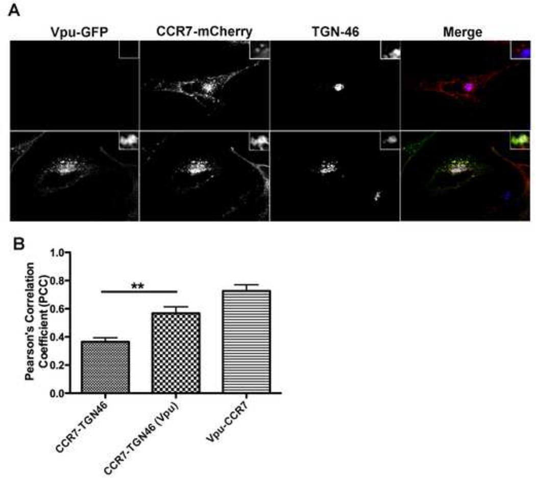
Figure 4. Vpu co-localizes with CCR7 within the trans-Golgi network.
A) HeLa cells were transiently transfected with either CCR7-mcherry alone (top row) or in combination with Vpu-GFP (bottom row). Twenty-four hours post transfection, cells were fixed, permeabilized and stained with a trans-Golgi network (TGN) specific antibody (TGN46). Images were acquired using a spinning disc confocal microscope. CCR7-mcherry (red); Vpu-GFP (green); TGN46 (blue).
B) Relative co-localization levels between CCR7-TGN46, Vpu-TGN46 or Vpu and CCR7 were quantified using Pearson’s correlation coefficient (PCC). Data is graphically depicted as mean +/− SEM PCC and is representative of ten individual cells where ** denotes a p value < 0.01.

