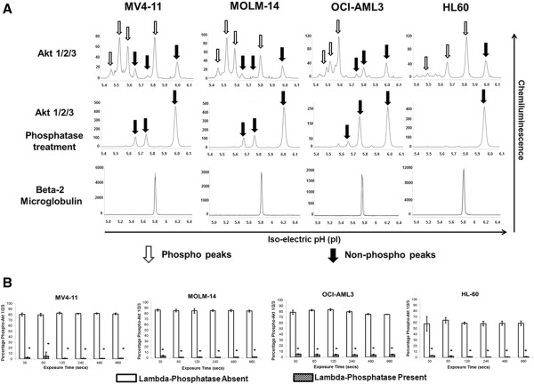
Figure 4.

Measurement of total and phosphorylated Akt 1/2/3 in AML cell lines. A) Electropherogram depicting levels of total Akt 1/2/3 in AML cell lines. AML cell lines MV4-11, MOLM-14, OCI-AML3 and HL60 were analyzed at baseline for activation of Akt. 80 ng of protein was used for analysis. β-2 Microglobulin was used as loading control. Total Akt antibody detects both phosphorylated and non-phosphorylated protein which is demonstrated on treatment with phosphatase. X-axis represents iso-electric pH and y-axis represents luminescence units. B) Different AML cell lines exhibit different levels of Akt phosphorylation as demonstrated using total Akt 1/2/3 antibody and the phosphorylation is diminished on treatment with lambda phosphatase. The experiments were performed in triplicate (*p < 0.05).
