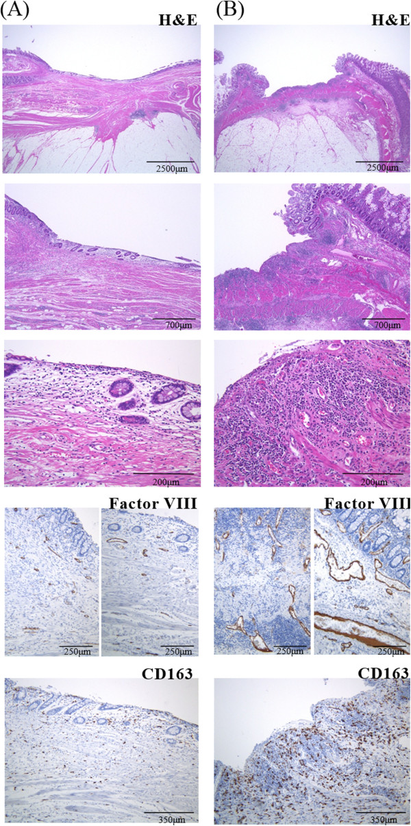
Figure 4.

Representative examples of pathological findings of the (A) “hypo-flow” and (B) “hyper-flow” surgical specimens. H&E staining revealed more active inflammation in “hyper-flow” (B) than in “hypo-flow” (A) cases. Vascular walls stained immunohistochemically using an anti-factor VIII antibody and macrophages stained with an anti-CD163 antibody were more prominent in “hyper-flow” (B) compared with “hypo-flow” (A) cases.
