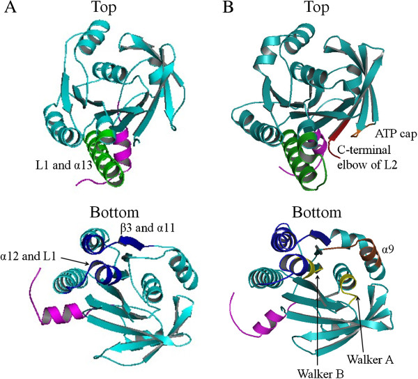
Figure 3.

Subdomain interactions at the interface of two neighboring ATPase domains in the PfuRadA ring structure (PDB: 1PZN) (A) and in the MvRadA filament structure (PDB: 1T4G) (B). In the ring structure, the interface of two neighboring ATPase domains is stabilized by L1 and α13 (in green) from one subunit (top) and α12, L1 and β3/α11 turn (in blue) from the adjacent subunit (bottom), while in the MvRadA filament structure, additional subdomains that interact at the interface between two adjacent ATPase core domains include ATP cap (in orange), the C-terminal elbow of L2 (in red) from one subunit (top), and Walker A, B motifs (in yellow), a part of α9 (in brown) from another subunit (bottom). The NTD is not shown for charity and the linker region between NTD and the core domain, including PM and SRM, is shown in pink.
