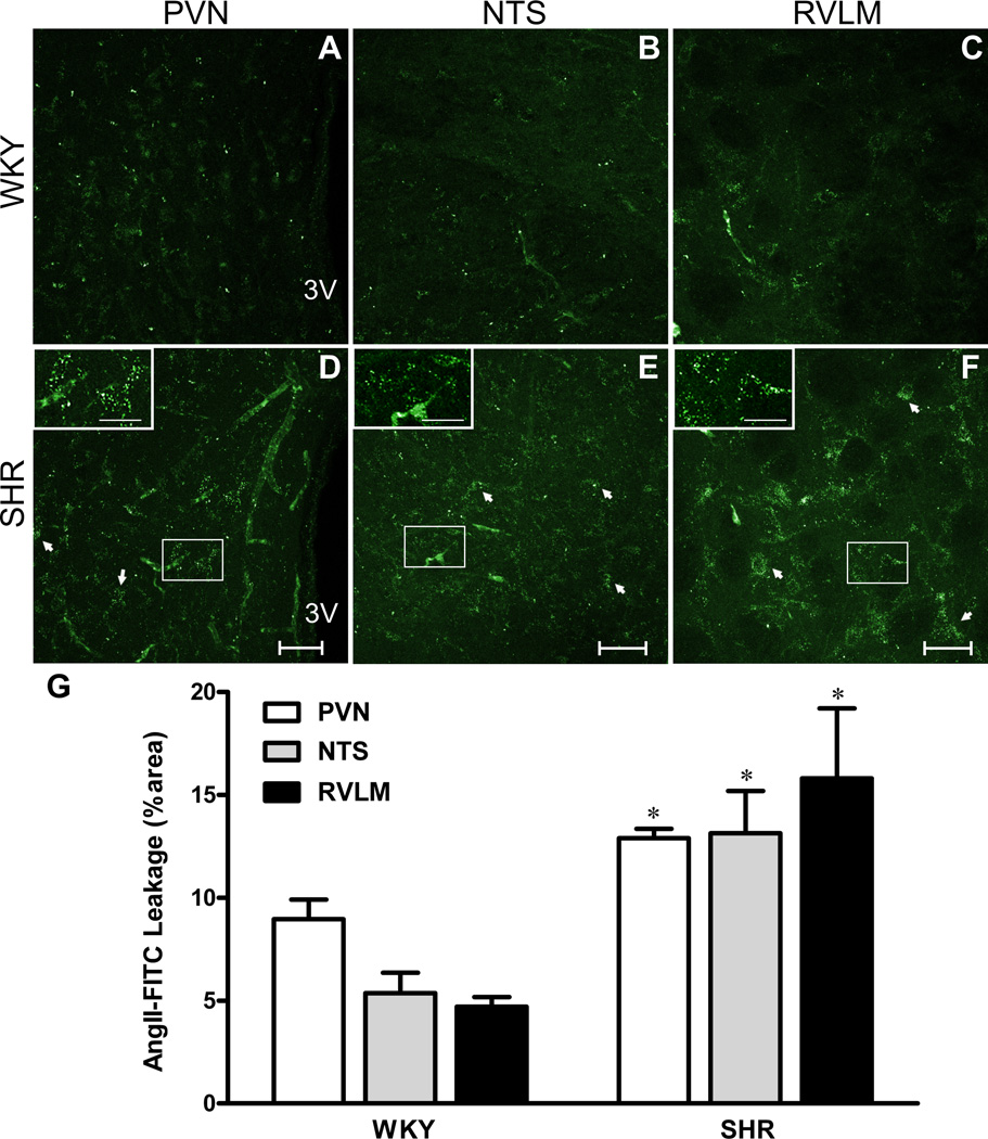
Figure 3. Circulating AngII leaks through the disrupted BBB in SHRs.
A–F, Intravascularly-delivered AngIIfluo within the PVN, NTS and RVLM of WKYs (A,B,C) and SHRs (D,E,F). Insets show respective squared areas at higher magnification. G, Mean extravasated AngIIfluo. *P<0.05 vs WKY, n=4/group. Scale bar: 50µm; insets: 25µm. 3V: third ventricle.

