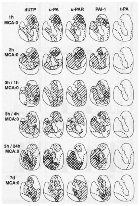
Fig. 3.
Time course of plasminogen activator responses to focal ischemia after middle cerebral artery occlusion (MCA:O), and MCA:O/ reperfusion in the basal ganglia of Papio anubis/cynocephalus.64 Nuclear deoxyuridine triphosphate (dUTP) incorporation defines regions of cellular injury in the basal ganglia. The distributions reflect the appearance by immunohistochemistry of individual plasminogen activator (PA) components associated with microvessels in the injured regions. Note that no upregulation of tissue plasminogen activator (t-PA) was observed. Each diagram represents the composite of three iterations (different subjects), with the hatched regions depicting 100% of the subjects displaying expression of the PA component of interest. Control tissues demonstrated no evidence of dUTP incorporation, u-PA, u-PAR, PAI-1, or t-PA expression.

